
As physiotherapists, it is important to understand the differential diagnosis that can cause knee pain to ensure that knee osteoarthritis is treated appropriately. In this page (and the previous page) you will learn more about the different conditions that can represent as knee pain to ensure you are aware of the various differential diagnosis.
Septic Arthritis
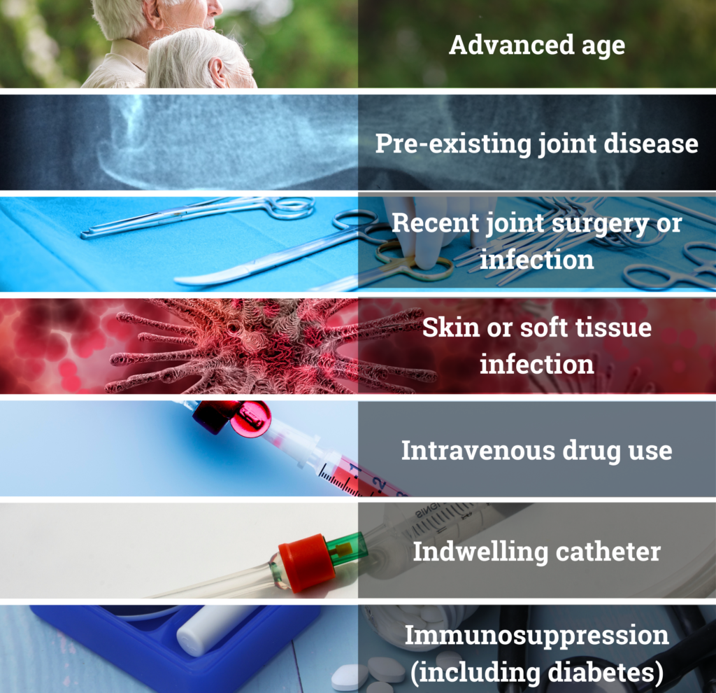 Septic arthritis is an infection in the joint. The risk factors for developing septic arthritis are found in the image to the right.
It normally occurs through an infection that is carried in the blood, but it can also be due to direct entry of bacteria into the joint through trauma, injection, or from adjacent tissues (e.g. osteomyelitis).
Patients usually have an acute, single swollen painful joint. Joint pain, swelling, warmth and restricted movement are present in approximately 85% of patients with septic arthritis.
- Most patients with septic arthritis are febrile, although older patients may be afebrile.
- Knee is the affected joint in over half of the cases.
The diagnosis is via synovial fluid analysis and culture (i.e. joint aspiration). Bloods and imaging should also be obtained; with imaging done to evaluate current bone and joint disease and not for diagnosing septic arthritis.
Differential diagnosis for septic arthritis include: gonococcal arthritis, lyme disease, tuberculosis, gout, pseudogout, reactive arthritis, OA flare up, acute traumatic arthritis (hemarthrosis associated with trauma to the joint)
Deep Vein Thrombosis
Deep vein thrombosis (DVT) can lead to life-threatening complications such as pulmonary emboli. Incidence of venous thrombo-embolism is estimated to be 1 per 1000 people per year, with approximately 2/3 of these being DVTs/ DVTs are slightly more common in males than females, and the incidence rises with increasing age.
Click on each of the info button to reveal the risk factors for DVT
History and clinical examination are not reliable ways of diagnosing DVT as patients can be asymptomatic.
Symptomatic patients may present with lower extremity pain, calf tenderness and asymmetric lower extremity swelling – however this is non-specific and suggest needs for further investigation rather than definitive diagnosis.
The Wells Score has been developed to reflect the risk of someone developing DVT.
(Sensitivity: 77-98%, Specificity 38-58%)
| Points | |
| -Active cancer (treatment ongoing or within previous 6 months or palliative | 1 |
| -Paralysis, paresis or recent plaster immobilization of the lower extremities | 1 |
| -Recent bedridden for 3 days or major surgery within 12 weeks requiring general or reginal anaesthesia | 1 |
| -Localised tenderness along the distribution of the deep veins | 1 |
| -Entire leg swollen | 1 |
| -Calf swelling 3cm > asymptomatic side (measured 10cm below tibial tuberosity) | 1 |
| -Pitting oedema limited to symptomatic leg | 1 |
| -Collateral superficial vein (nonvaricose) | 1 |
| -Previous DVT | 1 |
| -Alternative diagnosis as likely as or more likely than DVT | -2 |
| Score: DVT unlikely 1 ≤; DVT likely ≥ 2 | Total: |
DVT is normally diagnosed via either D-dimer, or venous ultrasound, or a combination of both.
Intermittent claudication (Neurogenic vs. vascular)
Neurogenic claudication relates to pain from a neural source – which is often associated with lumbar stenosis.
Vascular claudication is due to restricted blood flow.
Below is a table highlighting the difference between the two:
| Sign/symptom | Neurogenic | Vascular |
| Relief of pain | Sitting; bending forward; walking with a shopping trolley | Standing still |
| Proximal vs distal | Symptoms above knees | Symptoms below knees |
| Walking up hill | Better | Worse |
| Walking down hill | Worse | Better |
| Cycling | OK | Symptoms present |
| Walking distance | Variable | Fixed |
| Neurological symptoms e.g. P&N | Often present | Rarely present |
| Neurological signs | May be present | Absent |
| Pulse | Present | Absent |
| Skin | No changes | Atrophic |
Adapted from GP Notebook https://gpnotebook.com/en-au/simplepage.cfm?ID=x20170726144238191130
Cluster of symptoms is better in predicting whether it is neurogenic or vascular of origin.
Neurogenic symptoms are usually triggered with standing, relieved with sitting, located above the knees and has a positive shopping cart sign (walking is better when using a shopping trolley). If all these symptoms are present, there is strong evidence to suggest it is neurogenic in nature rather than vascular claudication
Vascular symptoms are usually relieved with standing alone, and are located below the knees.
Source:
 M. Nadeau, M. P. Rosas-Arellano, K. R. Gurr, S. I. Bailey, D. C. Taylor, R. Grewal, D. K. Lawlor, C. S. Bailey
Canadian Journal of Surgery (2013)
The reliability of differentiating neurogenic claudication from vascular claudication based on symptomatic presentation.
Reference:
Pigemented Villonodular Synovitis (or Tenosynovial Giant Cell Tumour)
 Pigemented villonodular synovitis (PVNS) otherwise known as tenosynovial giant cell tumour (TGCT) is a rare proliferative lesion of synovial tissue that causes thickening and overgrowth of the synovium. Some debate whether there is an inflammatory process or if it’s a benign neoplasm.
It is most common in the knee, but can be in other joints too. It can present with pain, swelling, arthritis and haemarthrosis. The disease itself is a hypervascular proliferative synovium and it may limit joint function and erode adjacent bone.
The main treatment is the resection of the synovium.
If you receive a referral with PVNS, refer on (orthopaedics, rheuamtology or both)
Spontaneous Osteonecrosis of the Knee (SONK)
 SONK is a category of osteonecrosis, with the precise etiology and pathology unknown. It is more common in females and should be suspected in individuals who are middle-aged to elderly, with sudden onset of spontaneous severe knee pain.
- It may be linked to subchondral insufficiency fractures and meniscal tears.
- X-rays are usually NAD initially. MRI is the preferred diagnostic imaging modality.
The management is also unclear. However, if you receive an imaging of a patient with SONK, or suspect that they may have it, your first action should be consulting the orthopaedic team.
- Usually the individual will be protected weight bearing and often need re-imaging to monitor changes.
Reactive arthritis
Reactive arthritis is joint pain and swelling triggered by an infection that is occurring in another part of the body, often 1-4 weeks after the infection . Most patients with reactive arthritis have
- Genitourinary or gastrointestinal signs and symptoms
- Eye symptoms (e.g. conjunctivitis)
- Skin or mucous membrane lesions.
It’s relatively rare and typically happen more to younger adults.
- In at least 50% of the patients, all symptoms resolve within 6 months, and most patients’ symptoms resolve within one year.
Synovial (Osteo)chondromatosis (SC)
Synovial (osteo)chondromatosis is a type of non cancerous tumour that is found in the lining of a joint. It is characterised by loose cartilaginous bodies that becomes osteochrondomatosis when these bodies ossify (70-95% of cases).
CT or MRI are recommended when SC is clinically suspected and radiographs either show a typical joint or show effusion only. However, when the loose bodies have ossified and becomes osteochondromatosis, these changes can be seen on x-rays too.
Erosions in the joint (because of the loose bodies) are less common in the knee in comparison to the hip.
There are thought to be two types:.
Primary SC (Reichel syndrome or Reichel-Jones-Henderson syndrome)is characterized by synovial metaplasia and proliferation that results in intra-articular cartilaginous looses bodies. Osteoarthritis occurs secondary to irritation in the joint from these loose bodies.
It is associated with preservation of articular cartilage initially.
Secondary SC is a disorder involving cartilaginous loose bodies because of pre-existing degenerative joint surface changes (such as trauma).
The differences between primary and secondary are:
- The intra-articular bodies are larger in secondary SC compared to primary SC
- There are less loose bodies in secondary SC compared to primary SC
- There bigger variations in size and shape of loose bodies in secondary SC in comparison to primary SC
- Secondary SC are often more severe on x-rays secondary SC because the loose bodies are due to osteoarthritic changes.